The word integumentary is derived from the latin word ‘integumentum’ meaning a covering. The integumentary system is the external covering of our body which includes skin, hair, nails and sweat glands. It is responsible for waste excretion and temperature regulation. It harbors the sensory receptors for pain, pressure as well as temperature. It makes up 16% of the total body mass and a surface area of 15-20 square feet. It is therefore considered as the largest organ system.The integumentary system is composed of the cutaneous membrane and accessory structures. The cutaneous membrane is further made up of the epidermis and dermis. The epidermis is the superficial epithelium while the dermis is the underlying connective tissue. The hair, nails and exocrine glands constitute the accessory structures.
Functions of the Integumentary System
1. Protection:
The chief function of the skin is protection of the underlying tissues from infection, mechanical, chemical and UV exposure as well as dehydration or desiccation.
2. Excretion:
Other than the kidneys, lungs, and liver; the skin also helps in the elimination of excretory wastes. It helps in the excretion of salts, water, and organic waste. The sweat and sebaceous glands in the skin can eliminate certain substances through their secretions. It thereby helps in maintenance of fluid and electrolyte balance.
3. Maintenance of body temperature:
The integumentary system helps in maintaining normal body temperature through conservation and radiation of heat with the assistance of sweat glands.
4. Synthesis of vitamin D3, which is essential for calcium metabolism:
A precursor of vitamin D is present in the blood, which is converted into the active form of vitamin D ( vitamin D3) only on exposure to sunlight. Vitamin D3 is utilized by kidneys for the synthesis of the hormone calcitriol, which is necessary for calcium absorption and thereby calcium metabolism. Thus, a vitamin D deficiency will reduce calcium absorption, which leads to weak bones.
5. Storage of nutrients i.e. Fats
6. Sensory detection:
The skin, along with its sensory receptors, helps us perceive the sensations of touch, pressure, pain, and temperature.
7. Non-verbal communication:
Human faces are very expressive because of the elasticity of the skin, and this is very helpful in non-verbal communication.
Layers of Skin
Epidermis, dermis, and hypodermis constitute the three parts of the skin, which we will see in detail below.

Epidermis:
The epidermis is the uppermost layer of the skin. Its upper portion is dead tissue filled with keratin that is a waxy protein. The lower layers have living cells that are replaced every 35-45 days. The epidermis is made up of different types of cells which have different functions.
The cells present are; stem cells, keratinocytes, melanocytes, Merkel cells and dendritic cells.
Stem cells are the undifferentiated cells that are found only in deepest layer.
Keratinocytes are the most abundant cells and they synthesize keratin. Melanocytes synthesize the pigment melanin.
Merkel cells are the touch receptors that are attached to nerve cells.
And finally, dendritic cells also known as Langerhans cells, are part of the immune system. These are macrophages that guard the skin against toxins, microbes and other pathogens which are able to penetrate the skin. On detection of such pathogens, they send an alert to the immune system.
Dermis (hide):
The dermis is the strong, flexible, connective tissue that provides strength and resilience to the skin. This gel-like matrix is made up of collagen, elastic and reticular fibers rich in nerves, receptors, blood vessels. It also has the hair follicles and sweat glands.
Subcutaneous Layer (hypodermis):
It is mainly the fat layer below the skin. It provides necessary insulation. Sensitivity to cold is more in the infants and elderly as they have thinner of the subcutaneous layer than adults.
Skin color
The color of our skin is decided by the pigments present. Both the epidermal pigments and blood pigments contribute to the skin color.
1. Epidermal Pigmentation:
Three different pigments are present in the epidermis: Melanin, Carotene, and Hemoglobin.
a) Melanin:
Melanin is synthesized from tyrosine, which is an amino acid. Every individual has a hundred melanocytes per mm2. The various racial shades are primarily due to the different kinds and amounts of melanin pigment. Sun exposure is also a factor that determines the amount of melanin pigment and thereby the skin shade. It also provides protection from UV rays to some extent.
b) Carotene:
It is a yellow-orange. It is obtained from the diet and not synthesized in the body as melanin. It is then converted into vitamin A, which is contained within the epithelium. It helps in maintenance of healthy epithelium as well as the photoreceptors.
c) Hemoglobin:
It is a red colored pigment present in the blood. Thus, the pigment that is present in the capillaries of skin contributes to its pigmentation.
2. Dermal Circulation:
Oxygenated blood contains hemoglobin pigment which is red in color. It gives a pink color to the skin. Upon vasodilation there is a flushing of the skin or the skin looks redder; while on vasoconstriction skin looks paler.
Abnormal Skin Color and Texture
Cyanosis – Skin appears bluish due to poor oxygenation
Erythema – Redness is seen which can be due to hypertension or inflammation
Pallor – Paleness due to low blood pressure or anemia
Jaundice- When the liver fails to excrete bile pigments, they start accumulating in the skin leading to yellowing of skin
Addison’s disease/Bronzing – An adrenal cortex hormone stimulates melanocytes to produce melanin. Whenever this hormone is produced in excess as in Addison’s disease, even melanin production is increased which gets deposited in the skin leading to bronzing.
Bruising /Hematoma – After trauma or injury, the escaped blood gets clotted leading to hematoma formation. Individuals with Vitamin C deficiency or hemophilia are prone to bruising
Albinism- It occurs due to a genetic mutation in melanin biosynthesis pathway leading to a lack of pigmentation in skin, hair and eyes. The person appears abnormally white and is very sensitive to light.
Vitiligo – It is an autoimmune disease wherein there is destruction of melanocytes
Skin Markings
Skin is marked by many lines, creases, and ridges.
1. Friction ridges: These are the markings on fingertips that are characteristic of primates. They allow easy manipulation of objects.
2. Flexion lines: On the outer surfaces of digits, palms, wrists, and elbows skin is very tightly bound to the underlying deep fascia leading to the formation of lines bending at these points. These lines are known as flexion lines.
3. Freckles: These are the flat melanised patches that vary with heredity or exposure to the sun.
4. Moles: These are elevated patches of melanized skin. They are considered as birth marks or beauty spots. Whenever hair is present within them, they are considered to be harmless.
Accessory structures
These structures though present within the dermal layer of skin, are derived from the epidermal layer during development.
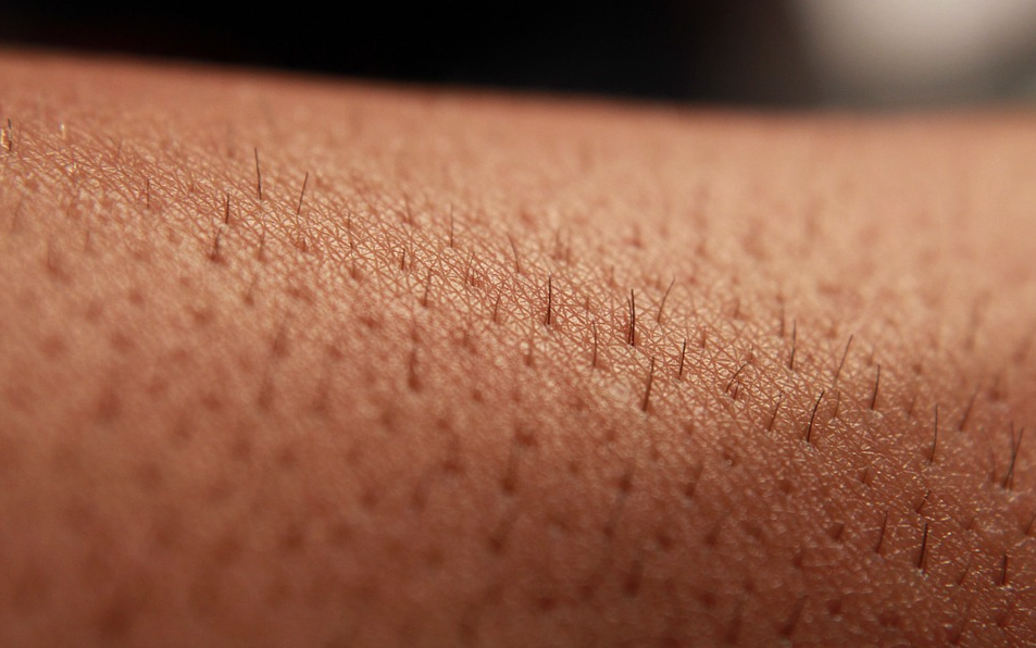
1. Hair

The human body has about 2.5 million hairs covering about 75% on our body. Hairs cover almost entire body except for lips, palms, soles, nipples and parts of external genitalia. After puberty hormones bring about the development of ‘hairy’ regions such as head, axillary and pubic areas. At birth, we have follicles for the hair we might develop with aging. The fastest growing tissues in the body include hair. Hair color is dependent upon the kind (yellow, rust, brown, and black) and the amount of melanin. The hair itself is dead, but is derived from live epidermal tissue.
2. Nails
These are actually a modified part of the epidermis. These are clear, hard and very thin. They are formed by dense packing of dead, scaly cells. Fingernails grow nearly 1 mm per week while toenails grow much more slowly. It has been found that addition of gelatin to diet does not have any effect on growth or hardness of nails. The appearance of nails can be of diagnostic value, for example spoonlike, flat, concave nails may be indicative of an iron deficiency, clubbed or swollen fingertips could be indicative of long-term hypoxemia resulting from congenital heart defects and/or emphysema etc.
3. Skin Glands: The skin is home to different types of glands
- Oil glands / Sebaceous Glands – These glands help in keeping the hair over skin soft and pliable especially on the face and scalp. There are about 2 or more glands per follicle. They are not present on the palms, soles or dorsal side of feet. They help reduce heat loss as fats are poor conductors of heat. They also help prevent evaporation of water. They become active at puberty when the acne secretes sebum which is a breakdown product of dead cells. Sebaceous glands eliminate certain substances like sterols, hydrocarbons, and waxes through sebum. This secretion provides a protective oily covering for the skin.
- Sweat Glands – There are about 3 million sweat glands in our body. They are most abundant on the palms, soles, forehead and armpits. An individual gland is like a tiny coiled tube which opens to the skin surface. Sweat glands help in maintaining the temperature and fluid-electrolyte balance. Sweat produced by the sweat glands is a watery fluid containing sodium chloride, small amounts of urea, lactic acid, etc. Though the primary function of sweat is to facilitate a cooling effect on the body surface, it also helps in the removal of some of the waste products mentioned above.
- Scent Glands – These are the modified sweat glands that produce scent or pheromones. They are much less common and are confined to the axillary and genital area. They respond especially to stress and sexual stimulation.
- Mammary Glands: These are modified sweat glands that produce milk.
- Ceruminous Glands: These are also the modified sweat glands present in the external ear canal. They secrete a waxy pigmented substance known as cerumin. It helps in trapping dust and other particles thereby protecting the internal ear.
Disorders of Skin
The skin being the outer envelope of our body is very susceptible. It can develop more than a thousand different ailments, the most common ones resulting from allergies or infections.
1) Allergies
Contact Dermatitis is an allergic response of skin to irritants such as poison ivy, metals etc.
2) Infections
Common viral infections of skin include cold sores, herpes simplex especially around lips and oral mucosa. Athlete’s foot is a common fungal infection of the skin. Bacterial infections are the most common, some of which are boils and carbuncles (inflammation of hair follicle and sebaceous glands mainly on the dorsal side of neck), Impetigo (Streptococcus infection) etc.
3) Diseases
Psoriasis and hypertrichosis are diseases that are fairly common. Psoriasis is a chronic, non-infectious skin disease wherein the skin becomes dry and scaly. Often there is a formation of pustules. There are many varieties of psoriasis. The associated genetic component is often triggered by trauma, infection, hormonal changes or stress. Hypertrichosis also commonly known as the human werewolves are cases wherein the patients show dense hair growth on faces and upper bodies due to malfunction of the gene on x chromosome. Alopecia areata is another condition wherein hair falls out in small round patches. It affects about 2% of the population (4.7M in the US). Hair loss is usually short-term and limited to a few patches. In very rare cases permanent loss of all body hair is seen.
4) Burns
Burns are categorized by the degree of penetration of heat into the skin layers.
- 1st-degree burns are where the skin is inflamed and the surface layer of skin is shed.
- 2nd-degree burns involve deeper injury with blister formation. There is fluid accumulation beneath the outer layers or epidermis.
- 3rd-degree burns involve the full thickness of the skin. Sometimes the destruction extends to involve even the subcutaneous tissues resulting in ulcerating wounds. There is typically catastrophic loss of fluids leading to dehydration and electrolyte imbalance. Also, susceptibility to infections is greatly increased. There is a slow recovery from cells of hair follicles if at all they survive; otherwise, healing takes place from margins of the wound. Autografts or cadaver skin may be required and prognosis depends upon the extent of the damage.
5) Skin Cancer
Skin cancer can be caused by excessive or chronic exposure to UV rays, X-rays or radiation. Most types of skin cancer progress slowly and can be successfully treated however a few forms are lethal.
a) Basal Cell Carcinoma: It is the least malignant and most common skin cancer. Middle third of the face is a common site of this cancer. There are tissue erosion and ulceration. 99% of these cancers are completely treated.
b) Squamous Cell Carcinoma: It is a type os skin cancer that is usually induced by sunlight.
c) Malignant Melanoma: It is the cancer of melanocytes i.e. the pigment producing cells. It is very rare but is fatal. It often begins with moles.
Age-Related Changes
Age-related changes often become noticeable by late 40s. With aging hair become thinner and start graying. There is atrophy of sebaceous glands leading to dryness of skin and hair. Skin becomes thinner and translucent. It also becomes looser and starts sagging. Dermis thins and becomes less elastic giving rise to wrinkles There is more bruising of skin due to increased fragility of blood vessels. Healing becomes delayed. Rosacea is seen, which are tiny dilated blood vessels, especially over nose and cheeks. Age spots are also visible due to the accumulation of pigment cells. Regulation of body temperature is less efficient due to loss of blood vessels and glands, increasing the vulnerability to hypothermia and heatstroke increases. Also, the production of Vitamin D3 declines and thus calcium absorption also declines, which leads to brittle bones.
- Photoaging: There is an acceleration of skin aging due to overexposure to the sun (UV rays). This accounts for 90% of the changes that people find to be of medical concern or cosmetically disagreeable.
Injury and Repair
The integumentary system can function independently of the nervous and endocrine systems to maintain its own homeostasis. Mesenchymal cells of the dermis can regenerate connective tissue while the germinative cells/ basal cells of the epidermis can regenerate epithelial tissue. Thus, in normal healthy individuals healing is very fast after any injury.
Summary
The integumentary system, considered as the largest organ of our body performs pertinent functions and is therefore of vital importance.