SKELETAL SYSTEM
The skeletal system is the system that provides a rigid framework to the body and thereby supports the soft tissues of the body. At birth there are about 270 bones in our body, but as growth and development completes many bones get fused together. Thus the total number of bones in the adult skeletal system is 206. The skeletal and muscular systems are intricately related to each other and together they are commonly called as the musculo-skeletal system. Bones with the help of joints and muscles bring about movements of the body.
Skeletal system composition
The skeletal system is primarily made up of bones. It also consists of the connective tissues associated with these bones. The connective tissues comprise of the cartilage, tendons as well as ligaments. Bone itself is also a type of connective tissue.
Based on their shape, there are four types of bone – long, short, flat, and irregular. Most of the bones of the upper and lower limbs are long bones. They are named so because they are longer than their width. Examples of short bones are the wrist and ankle bones. There are some bones that are flattened in shape like the skull bones, ribs, scapulae or shoulder blades, and the sternum. Irregular bones comprise the facial bones and vertebrae, which have unique shapes.

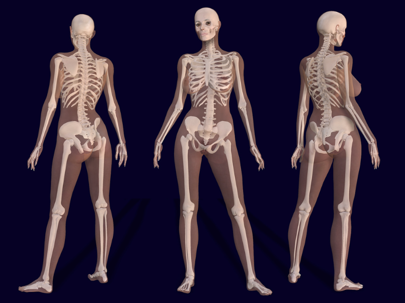
The skeletal system is primarily divided into the axial and the appendicular skeleton. Axial skeleton consists of the skull bones, bones of vertebral column, and the thoracic cage (sternum and ribs) while bones of the upper and lower extremities are included in the appendicular skeleton.
| BONES OF SKELETON | |||
| Axial skeleton (80 bones) | Appendicular skeleton (126 bones) | ||
| Skull (including hyoid bone) | 23 | Pectoral girdle | 4 |
| Ear ossicle | 6 | Upper extremities | 60 |
| Vertebral column | 26 | Pelvic girdle | 2 |
| Sternum and ribs | 25 | Lower extremities | 60 |
The skull is made up of 22 bones. It is composed of the cranial and facial bones. Cranium or the cranial bones are the bones that encase the brain by providing a rigid shielding outer layer. This outer layer is made up of 8 bones. There are 14 facial bones which shield the brain from the front side and form the facial structure. All the facial bones are firmly connected to form the basic constitution of the face except the mandible, which is not completely attached to the skull but is a freely movable joint.
Each middle ear has 3 small bones also called as the ear ossicles, incus, malleus and stapes.
The skull rests on two occipital condyles, which are located at the upper end of the vertebral column. Vertebral column is made up of 26 vertebrae extending from the base of the skull and can be felt along the back. It provides a rigid framework for the trunk portion of the body. The spinal cord is encased within the space at the center of the vertebrae along its entire length. The vertebral column has 7 cervical, 12 thoracic, 5 lumbar, 1 sacral and coccygeal vertebrae from the skull downwards. Basic function of the vertebral column is protection of the spinal cord and providing support to the head. The ribs as well as the musculature of back are also attached to the vertebral column..
Sternum is located at the front along midline of the chest. There are 12 pairs of ribs. Ribs are thin flat bones attached in the front to the sternum and at the back to the vertebral column. First seven pairs of ribs are known as true ribs as they are actually attached to the sternum and vertebral column. However the 8th, 9th and 10th pairs of ribs are not directly attached to the sternum but are joint to the 7th rib with hyaline cartilage. These are known as false ribs. Last 2 pairs of ribs i.e. 11th and 12th ribs are not connected in the front and hence are known as the floating ribs. Thoracic vertebrae, ribs and sternum are collectively called the rib cage.
Limb bones and girdles together form the appendicular skeleton. Each limb consists of 30 bones. Humerus, radius and ulna are the long bones of upper limbs. There are 8 wrist bones or carpals, 5 palm bones or metacarpals and 14 digits or phalanges. In the lower limbs, femur, tibia and fibula are the long bones of which femur or thigh bone is the longest. The ankle is made up of 7 bones, known as tarsals. Further there are 5 metatarsals and 14 digits or phalanges. There is a peculiar cup shaped bone covering the knee in the front known as the patella. The upper and the lower limbs articulate freely because of the fixed pectoral and pelvic girdles to which they are attached respectively.
Two symmetrical halves make up each girdle. Pectoral girdle (to which the upper limbs are attached) consists of a clavicle and a scapula. Scapula is a large triangular flat bone located at the back bilaterally and a part of the scapula articulates with the head of the humerus to form the shoulder joint. Clavicle is a long slender bone known as the collar bone and forms the front part of the pectoral girdle. Pelvic girdle supports the lower limbs and is made up of two coxal bones. Ilium, ischium and pubis are the three bones that form the pelvic girdle and also provide articulation for the thigh bone or femur. The two halves of the pelvic girdle join in the front as the pubic symphysis which is made up of fibrous cartilage.
Bone Repair occurs naturally and essentially whenever a bone is broken. When a bone is injured blood vessels in the bone are also damaged leading to bleeding and clot formation in the damaged area. Two to three days later a fibrous network of connective tissue is prepared between the broken bones by the blood vessels and cells from surrounding tissues that begin to invade the clot.
This newly formed connective tissue holds the bone fragments together thus filling the gap between bones. Islets of cartilage are also produced by some cells in the fibrous network. This is known as callus. Further the bone forming cells or osteoblasts, enter the callus and gradually replace it with bone, which completes within 4–6 weeks after injury.
Immobilization of the bone is of utmost importance at this time because the new matrix formed is very fragile and is likely to re-fracture. Subsequently, there is remodelling of the spongy bone to form the compact. This completes the repair. Immobilization is critical but should not be extended for a very long span as it could lead to muscle wasting and reduced muscle strength. Thus modern treatment aims to balance immobilization with adequate amount of exercise. This prevents muscle wasting and at the same time the size and strength of bones is maintained. This is accomplished by use of “walking casts and restricting the amount of time a cast is left on the patient. This allows some stress on the bone and some movement thus helping in maintaining the joint mobility. Complete healing of fractured bone may require many months.
Muscle is a specialized tissue that contributes to nearly 40-50 per cent of the body weight in a human adult. Muscles are primarily classified as skeletal visceral and cardiac. Skeletal muscles are those associated with the bones of the body. They are called as the voluntary muscles because their activities can be voluntarily controlled. They are mainly involved in body posture changes and locomotory movements.
Joint or articulation, is where two bones come together. Joints are necessary for movements involving the skeletal parts of the body. This mainly involves the locomotory actions. Joints help in forming an attachment between two bones, or between bones and cartilages. This attachment is such that is allows a specific range of movement. Muscles help in bringing about the movement through joints, where the joint acts as a fulcrum.
Three types of joints are formed between bones or between bone and cartilage – fibrous, cartilaginous and synovial.
Fibrous joints allow very limited movement. This type of joint is shown by the flat skull bones which fuse end-to-end with the help of dense fibrous connective tissues in the form of sutures, to form the cranium.
Cartilaginous joints are joints wherein the bones are joined together with the help of cartilages. They usually allow limited movement. An example is the joint between the vertebrae of the vertebral column wherein limited movement is permitted.
Synovial joints are the ones which allow considerable movements and therefore, play a significant role in locomotion. They are by the presence of a fluid filled synovial cavity between the articulating surfaces of the two bones thus allowing considerable movement. These joints collectively assist in locomotion and various other movements. Ball and socket joint (between humerus and pectoral girdle), hinge joint (knee joint), pivot joint (between atlas and axis), gliding joint (between the carpals) and saddle joint (between carpal and metacarpal of thumb) are examples of synovial joints.
Types of Movement
Joints are capable of different movements and these movements are related to its structure. Thus some joints can perform movements in different directions while some are permitted only limited movement. The different types of movements are flexion, extension, abduction, adduction, pronation, supination, elevation, depression, circumduction, rotation.

Some other movements like eversion, inversion, protraction and retraction are also used. Of these flexion and extension are most commonly performed movements. Flexion literally means to bend and extension means to straighten. Abduction is an action of taking away from the midline of body while adduction is movement towards the body midline. For example, movement of legs away from body midline as done while performing ‘jumping jacks’ is abduction, and adduction is when the legs are brought back together.
Pronation is when on rotation of forearm the palm faces downwards and supination is rotation of forearm so that the palm faces upwards. Eversion is when the bottom of the foot faces outwards and inversion when foot faces inwards. When a structure turns around its long axis such as when shaking the head to say ‘no’ it is rotation. Circumduction is the free movement such as the shoulder joint. Usually locomotion is brought about by a combination of movements.

Functions of the Skeletal System
- Bones: Bones are very strong and are thus apt for bearing weight. They provide a framework which gives a shape/ form to the body. Cartilage is flexible as compared to bones but yet it adequately supports structures like nose, external ear and trachea. Support is also provided by fibrous bands known as ligaments which are directly attached to bones. They thus help to hold the bones together.
- Protection of organs: Bones being very hard and strong provide excellent protection to the organs that it surrounds. Brain is a very well protected by the skull. Similarly heart and lungs are enclosed within the rib cage.
- Movement: Bones help in bringing about the movements with the assistance of skeletal muscles. Skeletal muscles are attached to the bones by tendons. Whenever the skeletal muscles contract, the bones produce body movements. Movement between two bones is because of joints. Ligaments supervise the range of movement and prevent excessive movement.
- Calcium and phosphorus storage: Bone stores certain minerals from blood, mainly calcium and phosphorus. Whenever the levels of these minerals in the blood decreases, bone releases these minerals from its store into the blood so that the levels in blood get back to normal. Adipose tissue is also stored within bone cavities. Lipids are also stored in the bone in the form of adipose tissue and are released into blood when necessary. Lipids are utilised as an energy source by other tissues.
- Production of blood cells: Bone is filled with bone marrow. The red bone marrow synthesizes the red blood cells, white blood cells and platelets.
Disorders of muscular and skeletal system
Like all human structures, bones and joints can also be affected by disease/s.
- Fracture of bones: There is fracture of bone whenever it is subjected to excessive forces. This leads to formation of a gap between the bones. If the fractured ends are not displaced only a cast for immobilization is enough for healing however if they are displaced to a great extent then an orthopaedic surgeon may align the bones under anaesthesia to prevent deformity.
- Sprain: If the ligamentous structures around a joint are injured it causes sprain. Here the anatomic relations are maintained.
- Dislocation of joints: A dislocation is when the end of one bone is pulled out of the socket in a joint mainly the ball-and-socket, ellipsoid, or pivotal joint. There is complete loss of joint integrity and loss of anatomic relationship.
- Osteogenesis imperfect: It is defective bone formation that is related to gene mutations encoding collagen type I. It affects bones as well as connective tissue. Clinical symptoms depend on the severity of the disease.
- Osteoarthritis: It is a degenerative joint disease involving the entire synovial joint. It is likely to develop whenever the joint is under stress and mostly affects weight bearing joints like hips, knees, vertebrae. It is an active disease process wherein there is joint destruction and aberrant repair because of alterations in cellular function. Genetics, nutrition, weight control, bone density, estrogen use, local biomechanical factors, previous injury are the factors which contribute to its etiology. Its prevalence increases with age.
- Rheumatoid arthritis: It is an inflammation of joints primarily involving the synovial joints. Secondary to inflammation pannus develops and covers the articular surfaces like a sheet. Pannus is rich in inflammatory cells which secrete lytic enzymes. Almost 1% of world population is affected by rheumatoid arthritis and it is more common in women. Its etiology is unknown however contributing factors are chronic, systemic inflammatory disease, genetic and familial predisposition, environmental triggers.
- Gout: Accumulation of uric acid crystals within the joint leading to inflammation of joints and therefore reduced and painful movements.
- Osteoporosis: Age-related disorder characterized by decreased total bone mass density (BMD) and increased susceptibility to fractures. Reduction in the levels of estrogen is a common cause.
- Osteomalacia: It is the softening of the bones caused by deficiency of Vitamin D or that of phosphate metabolism which leads to inadequate mineralization of bone matrix. It usually starts with diffuse, generalized aching and fatigue. However bone deformities like bow legs and thoracic kyphosis are seen as the disease progresses.
- Tumors of bone: Osteosarcoma, Chondrosarcoma and Ewings sarcoma are the tumors that affect the skeletal system.
- Osteomyelitis: Osteomyelitis is the inflammation of bones caused by infectious organisms mainly bacteria. Bacteria enter the bones through blood circulation as bones are porous. The infection thus spreads easily and rapidly. Further there is pus formation and in long standing cases it spreads to adjacent structures too. Body tries to wall off the infection by formation of reactive bone.
_______________
Featured image courtesy Bernard Ungerer